ntvanessa
New
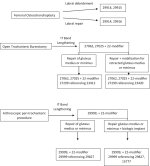
I'm struggling with this. Hope someone can help solve this puzzle. My supervisor showed me an article from NLM published in 2021 for hip in which they suggested the coding as attached for hip procedure. I feel that just billing 29999 for both arthroscopic IT release and bursectomy the provider won't get all the reimbursement for the work he did. Copy of the operative report below for review. Appreciate the help!!!
60 mL of normal saline was then injected directly over the IT band in the midportion of the trochanter to displace the soft tissues. Distal incision was made approximately 2 cm below the vastus ridge. In a proximal incision was made approximately 2 cm above the tip of the greater trochanter. The soft tissue was then bluntly dissected off the IT band using the camera trocar. Camera was then placed and the shaver was then used to visualize the IT band by removing a small amount of the soft tissue overlying it. Spinal needle was then placed in the mid aspect of the greater trochanter. This was utilized as a guide to split the IT band. The IT band was then split with an ArthroCare wand just to the posterior aspect of the midportion of the trochanter. 2 transverse splits were then made one anteriorly and one posteriorly in the standard fashion approximately 1 cm long in each direction. The greater trochanteric bursa was noted to be very thickened. This was then split using a frequency wand being careful not to injure the vastus lateralis fascia or the abductor tendons. The motorized shaver was then used to debride out the remaining greater trochanteric bursa. Care was taken not to go to posterior in the region of the sciatic nerve. Once a good decompression and release of the IT band was obtained the camera was then switched to the opposite portal and the release was further checked and noted to be sufficient. Final pictures were taken.

60 mL of normal saline was then injected directly over the IT band in the midportion of the trochanter to displace the soft tissues. Distal incision was made approximately 2 cm below the vastus ridge. In a proximal incision was made approximately 2 cm above the tip of the greater trochanter. The soft tissue was then bluntly dissected off the IT band using the camera trocar. Camera was then placed and the shaver was then used to visualize the IT band by removing a small amount of the soft tissue overlying it. Spinal needle was then placed in the mid aspect of the greater trochanter. This was utilized as a guide to split the IT band. The IT band was then split with an ArthroCare wand just to the posterior aspect of the midportion of the trochanter. 2 transverse splits were then made one anteriorly and one posteriorly in the standard fashion approximately 1 cm long in each direction. The greater trochanteric bursa was noted to be very thickened. This was then split using a frequency wand being careful not to injure the vastus lateralis fascia or the abductor tendons. The motorized shaver was then used to debride out the remaining greater trochanteric bursa. Care was taken not to go to posterior in the region of the sciatic nerve. Once a good decompression and release of the IT band was obtained the camera was then switched to the opposite portal and the release was further checked and noted to be sufficient. Final pictures were taken.
