Radiology Coding Alert
Precisely Code Colles’, Smith’s, and Barton’s Fractures With This Guide
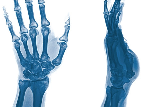
Hint: Bone fragment positioning determines the diagnosis. Of the assortment of wrist fractures in ICD-10-CM, three types stand out with unique names — Colles’, Smith’s, and Barton’s fractures. Knowing the difference between these three fractures will help you assign the correct diagnosis code. Start here: Under the S52.5- (Fracture of lower end of radius) code subcategory in the ICD-10-CM code set’s tabular list, you’ll find several subcategories related to distal end (lower end) radius (wrist) fractures. Confidently Code a Colles’ Fracture Diagnosis A Colles’ fracture “is the most common fracture and results in dorsal displacement, which causes the bone fragments to bend toward the back of the hand,” says Grabiela Juarez, COC, CPC, CPMA, Approved Instructor, revenue cycle specialist of Sceptre Management and owner of Medical Coding Vida Academy in Salt Lake City, Utah. In a Colles’ fracture, the dorsal displacement can result in a “dinner fork deformity,” where the patient’s wrist resembles an overturned dinner fork. A patient who suffers a Colles’ fracture may experience the injury when they extend their hand to catch themselves during a fall. It can also occur with patients experiencing trauma in a car accident, while playing contact sports, or when skiing, skating, horseback riding, or bike riding. The break causes severe pain and is a very serious injury. The codes listed in the ICD-10-CM code set for a Colles’ fracture include: Scenario: A patient presents to the emergency department (ED) with severe wrist pain after falling off their bicycle. After a physical exam, the physician notes the patient’s dinner fork deformity and orders lateral and anteroposterior (AP) X-rays of the left wrist. The radiologist captures the views of the patient’s wrist and reports their findings. The physician diagnoses the patient with a closed Colles’ fracture of the left wrist. For this scenario, the radiologist will assign 73100 (Radiologic examination, wrist; 2 views) for the X-rays of the patient’s wrist. Append modifier 26 (Professional component) if you’re reporting only the radiologist’s professional work. You’ll then assign S52.532A (Colles’ fracture of left radius, initial encounter for closed fracture) for the patient’s diagnosis. Additionally, you also may assign an external cause of morbidity code from Chapter 20: External Causes of Morbidity (V00-Y99). For this scenario, you’ll assign V18.0XXA (Pedal cycle driver injured in noncollision transport accident in nontraffic accident, initial encounter) to report the patient suffered the injury after falling off their bicycle. While there are no national mandates for reporting external cause of morbidity codes, there may be local or state requirements and particular payers may require them for liability purposes. Study How to Code a Smith’s Fracture In a Smith’s fracture, there is a forward displacement of the distal end of the radius. This volar, or forward, displacement causes the bone fragment to bend toward the palm side of the hand. Due to the positioning of the bone fragment, a Smith’s fracture is also known as a reverse Colles’ fracture. A Smith’s fracture occurs when the patient falls or experiences another trauma while the wrist is flexed or bent. While Smith’s fractures are rare, they are more common in young males as well as elderly females diagnosed with osteoporosis who experience low-impact falls. The physician may look for the “garden spade deformity” when examining the patient’s wrist. When the injury occurs, the fracture gives the appearance that the patient’s hand is being pulled toward the body. A radiologist will capture X-rays to check for a fracture, and may need to perform computed tomography (CT) or magnetic resonance imaging (MRI) scans to rule out or confirm damage to muscles, tendons, and ligaments. In the ICD-10-CM code set tabular list, you’ll find Smith’s fracture codes under the following: Scenario: A 75-year-old female patient arrives at an urgent care clinic following a fall in her home. The patient has a history of osteoporosis and tripped on an area rug while carrying a planter. The physician assistant (PA) performed a physical exam, noted the patient’s wrist appeared to have a garden spade deformity, and ordered X-rays to confirm a fracture. The radiologist captured lateral, AP, and oblique views of the patient’s right wrist. Following the X-rays, the radiologist listed their findings as a Goyrand fracture. In this scenario, you’ll select S52.541A (Smith’s fracture of right radius, initial encounter for closed fracture) for the patient’s injury diagnosis. “Named after the French physician Jean-Gaspard-Blaise Goyrand, Smith’s fractures are often referred to as Goyrand fractures in French literature,” Juarez says. Additionally, you’ll assign 73110 (… complete, minimum of 3 views) to report the X-rays. Boost Your Barton’s Fracture Coding Knowledge A Barton’s fracture is a dislocated wrist fracture that occurs when the distal end of the radius breaks near the base of the thumb and the fracture extends into the wrist joint. In this intra-articular fracture, if the bone fragment moves forward then the patient experiences a volar (anterior) Barton’s fracture, but if the bone fragment moves backward then the patient experiences a dorsal (posterior) Barton’s fracture. Either way, you’ll choose the appropriate code from the S52.56- (Barton’s fracture) subcategory, such as: Scenario: A patient is referred from their primary care physician to your radiology practice for X-rays of the patient’s left wrist. While playing soccer, the patient fell and hurt their wrist upon hitting the ground. After capturing posteroanterior (PA) and lateral X-ray views of the patient’s left wrist, the radiologist documents their findings of a closed Barton’s fracture of the left radius. In this scenario, you’ll assign S52.562A (Barton’s fracture of left radius, initial encounter for closed fracture) to report the patient’s diagnosis. Bottom line: Staying aware of the direction the fractured bone is pointing will help you remember which fracture to code. “The difference between the three is that the broken piece of the wrist bone points or is displaced backward in a Colles’ fracture, forward in a Smith’s fracture, and Barton’s fracture includes an intra-articular [joint] component,” says Taylor Berrena, COC, CEMC, CFPC, coder II at Cooper University Health Care in Yorktown, Virginia.


Related Articles
Radiology Coding Alert
- Ultrasounds:
Know What A- and B-Scans Mean for Ophthalmic Ultrasounds
Do you need to permanently store images? Find out. When you examine the code descriptors [...] - ICD-10-CM:
Precisely Code Colles’, Smith’s, and Barton’s Fractures With This Guide
Hint: Bone fragment positioning determines the diagnosis. Of the assortment of wrist fractures in ICD-10-CM, [...] - Industry Notes:
Contrast Supply Shortage Leaves Radiology Practices Scrambling
Will practices be able to change previously authorized orders? Radiology practices across the U.S. have [...] - You Be the Coder:
Get Specific When Selecting X-ray Codes of Extremities
Question: A 65-year-old patient with a history of osteoporosis visited our urgent care clinic complaining of [...] - Reader Questions:
Make Sure You’re Using Updated Codes to Report X-rays
Question: Our radiologist captured bilateral anteroposterior (AP), posteroanterior (PA), and lateral views of a patient’s hips [...] - Reader Questions:
Can You Report a Bulging Disc as Displaced?
Question: A patient was referred to our radiology practice for X-rays of their lumbosacral region. The [...] - Reader Questions:
Know When to Separately Report Fluoroscopic Guidance
Question: A radiologist in our practice performed fluoroscopic guidance for a diagnostic lumbar spine puncture of [...]


