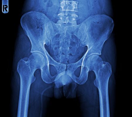
Do you need multiple codes for DXA and VFA scans? Radiologists use dual-energy X-ray absorptiometry (DXA) scans to measure a patient’s bone density to diagnose several conditions. As a radiology coder, it’s up to you to ensure the DXA code you choose matches the body area scanned to ensure proper reimbursement. Dive into DXA scans and learn how to choose the correct codes for each procedure. Diagnose DXA Scan Procedures A DXA scan is a noninvasive imaging method physicians use to measure the mineral amount in the patient’s bones. This measurement is referred to as the bone mineral density (BMD). As a patient grows older, the body naturally loses its ability to generate new bone material to replace bone loss. During the procedure, the provider aims two X-ray beams with different energy levels at the patient’s bones. The scanner calculates the BMD by gauging the absorption of each beam by the patient’s bone. The physician then interprets the absorptiometry — if the absorption is high, then the bone is dense and strong. Providers use DXA scans to determine a patient’s risk of fracture as well as diagnose bone-related health problems like osteoporosis and osteopenia, the latter of which occurs as the bones lose mineral mass and become thinner while the patient grows older. Documentation: Without proper documentation, your DXA test claim will likely be denied. “A DXA test must include the report with assessment as it relates to the medical history of the patient and interpretation. The report must be separate and distinct from an evaluation and management (E/M) visit,” says Grabiela Juarez, CPC, CPB, CPMA, COC, AAPC-Approved Instructor, billing manager at Summit Foot and Ankle and owner of Medical Coding Vida Academy in Salt Lake City, Utah. Additionally, the report needs to show the T and Z scores according to Juarez. A T score evaluates the strength of the bone and the estimated risk of the patient experiencing a fracture, whereas a Z score compares the patient’s bone density with that of an average person of the same age and sex. Examine the following scenarios to learn how to code different DXA scans correctly. Analyze This Axial Skeleton DXA Scan Scenario: A 75-year-old female patient presents to a radiology practice for a DXA scan. The radiologist positions the DXA scanner over the patient and scans the patient’s hips and pelvis. After interpreting the results, the radiologist documents their findings as osteoporosis. To report the DXA scan for this encounter, you’ll assign 77080 (Dual-energy X-ray absorptiometry (DXA), bone density study, 1 or more sites; axial skeleton (eg, hips, pelvis, spine)). The provider scanned the patient’s hips and pelvis, which are part of the axial skeleton. The axial skeleton is composed of the bones that lie in the central core of the human body. These 80 bones include the skull, neck, back, and ribcage bones. You’ll assign the code only once regardless of how many axial skeleton sites the provider scans during the procedure. Recognize This Forearm DXA Scan Scenario: A 55-year-old patient presents to a radiology practice for a DXA scan after recently experiencing a left forearm fracture. The patient fractured their arm in a minor fall at home, and their primary care physician (PCP) wanted to assess the patient’s risk for future fractures. The radiologist positioned the scanner and performed the procedure on the patient’s arms and legs. The radiologist then reviewed the results and documented their findings of osteoporosis. In this scenario, you’ll assign 77081 (… appendicular skeleton (peripheral) (eg, radius, wrist, heel)) to report the procedure. The appendicular skeleton consists of the body’s upper and lower extremities, such as the arms and the legs. Use This DXA Procedure for Suspected Spinal Fracture Assessment Scenario: A 65-year-old female patient with a history of osteoporosis visited an outpatient radiology facility. She had been experiencing severe back pain for several weeks. The patient’s PCP suspected a vertebral fracture due to her medical history and symptoms and decided to order a vertebral fracture assessment (VFA). The radiologist positions the patient positioned on the DXA machine and performs a VFA scan. After reviewing the images, the radiologist finds evidence of a vertebral fracture. In the CPT® code set Index, you’ll find two codes listed under Dual-Energy X-ray Absorptiometry (DXA) > Vertebral Fracture, but you need only one of those codes to report this encounter. You’ll assign 77086 (Vertebral fracture assessment via dual-energy X-ray absorptiometry (DXA)) to report the radiologist’s procedure in this scenario. Procedure definition: The VFA is a low-dose X-ray exam performed on a DXA scanner to check the patient’s spine for bone breaks in the vertebrae. Verify an Axial DXA Scan With a Vertebral Fracture Assessment Scenario: An 85-year-old patient visits a radiology practice for a DXA scan. The patient’s PCP referred the patient for the procedure after recognizing the patient has lost 1.5 inches of height at their most recent annual physical. The radiologist performed a DXA scan of the axial skeleton and a VFA. After reviewing the results, the radiologist documented their findings. Returning to the CPT® code set Index, you’ll select the other available VFA code to report this procedure. Assign 77085 (… axial skeleton (eg, hips, pelvis, spine), including vertebral fracture assessment) on your claim to report the DXA scan and VFA procedure. Code 77085 covers the DXA scan of the axial skeleton as well as the VFA. On the other hand, if the provider performs a DXA scan of the patient’s appendicular skeleton and VFA, you’ll assign 77081 and 77086 to report the procedures.