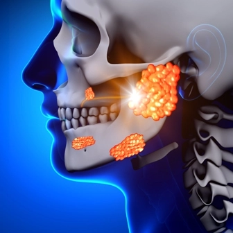
Switch to K11.5 For Reporting Sialolithiasis in ICD-10

If your oral surgeon identifies the diagnosis as sialolithiasis, you will report the same ICD-10 code irrespective of which of the salivary glands or their duct is affected by the stone formation. Your reporting of the condition in ICD-10 is very similar to the way in which you reported the condition using ICD-9 code sets.
ICD-9: When your oral surgeon arrives at a diagnosis of sialolithiasis, you report the condition with the ICD-9 code, 527.5 (Sialolithiasis). You report the same diagnosis code if your clinician’s diagnosis is calculus of the salivary gland or duct. The same diagnosis code can be used if your clinician diagnoses the patient with stone in the salivary gland or the duct. You also use 527.5 if your clinician diagnoses the patient with sialodocholithiasis.
Caveat: You cannot use 527.5 if your clinician’s diagnosis is sialoadenitis. You use have to use 527.2 instead of using 527.5. You also cannot use 527.5 if your clinician diagnoses the patient with disturbance in salivary secretion. In such a case, you will use 527.8 instead of 527.5.
Note: There are no separate ICD-9 codes to specify which of the salivary glands (submandibular, parotid or sublingual) or their ducts are affected by the calculus formation. Since there are no specific codes to identify the appropriate salivary gland or its duct, you use the same code for the diagnosis irrespective of which of the glands (or ducts) are involved.
ICD-10: When you begin using ICD-10 codes instead of ICD-9, a diagnosis of sialolithiasis that you would report with 527.5 crosswalks to K11.5 (Sialolithiasis). As with ICD-9, you use the same ICD-10 code if your clinician diagnoses the patient with calculus of the salivary gland or duct. You also use K11.5 when the diagnosis is mentioned as stone in the salivary gland or duct.
Reminder: As with ICD-9, you cannot use K11.5 for a diagnosis of sialoadenitis. You report this condition with one of the codes from the range, K11.20-K11.23 depending on chronicity of the condition. For cases of disturbances in salivary secretion, you cannot use K11.5. Instead, you will have to report this diagnosis with K11.7 (Disturbances of salivary secretion).
As with ICD-9 codes, there has been no specificity introduced to identify which of the salivary glands (or their ducts) are affected due to the calculus formation. So, in ICD-10 too, you will only report one code irrespective of the gland or the duct in which the calculus or calculi are formed.
Brush up on These Basics Briefly
Documentation spotlight: A few of the common findings that your clinician will record when he arrives at a diagnosis of sialolithiasis will include pain in the cheek or the area below the tongue, and swelling and distension in the affected area. The patient might also complain of fever and chills and disturbances with swallowing of food. Some patients might also have complaints of dryness of the mouth, difficulty in breathing and pain in the area of the lymph nodes. Your clinician will record a thorough history of the patient that includes onset and duration of symptoms, whether the symptoms are occurring for the first time or it is recurrent, recent dental history, medical history, drug allergies, immunization history and history of any other recent surgical or radiation treatments.
Upon examination, your clinician might note the presence of a swelling in the affected area. Your clinician will also palpate the affected gland to check if he can detect any calculi present. Your clinician will also examine the opening of the salivary gland duct to check if there is any outflow of saliva from the duct and also to note if there is any purulent discharge from the ductal outlet. Your clinician will also examine the adjacent areas to check for any changes in the normal appearance. Your surgeon will also check the lymph nodes to see if there are any signs of lymphadenopathy.
Tests: If your surgeon suspects the patient to be suffering from sialolithiasis, he will order for certain lab tests and imaging studies to confirm the diagnosis. He will order for routine complete blood count with differential count along with electrolyte studies to rule out the presence of any infections in the salivary gland or in the duct.
Your surgeon will also want to order imaging studies such as plain radiography using different views or sialography (which involves injecting a water-soluble medium such as meglumine diatrizoate into the ductal opening and then performing plain radiographs in different views to check if the duct has any blockage due to the presence of calculi.
Your clinician might also order for other imaging studies such as ultrasonography, CT or an MRI scan to arrive at the diagnosis of sialolithiasis although an MRI scan is not very helpful in identifying the calculi.
Your surgeon will initially try to manage the condition by using a warm compress, through hydration or by gentle massage of the gland. If the condition becomes recurrent, he will perform surgery to help the patient obtain relief. If the calculus is present near the ductal opening, he will cannulate and dilate the ductal opening using an intraoral approach and then reach out to the stone in the duct and remove it. If the stone is located deep inside or if multiple calculi are present, your clinician might opt to excise the entire gland itself along with the associated duct.


