Neurosurgery Coding Alert
Less-Invasive Stereotactic Surgery Requires Special Coding Skills
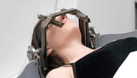
You’ll choose 1 of 2 codes when surgeon operates on intracranial lesions. When your neurosurgeon performs a stereotactic procedure to address intracranial lesions, they’re usually trying to avoid a more invasive surgery or procedure, saving the patient recovery time and cutting down on the surgical risks that could be incurred. “Stereotactic surgery is not an ‘otomy’ or an ‘ectomy,’” said Oby Egbunike, COC, CPC, CRC, AAPC Approved Instructor, during her HEALTHCON 2022 Denver presentation “Stereotactic Procedures for Diagnosis of Intracranial Lesions.” To code for these surgeries properly, you’ll need to know several facts regarding head frames, CPT® codes, and imaging. Read on for the info you’ll need to correctly code your stereotactic radiosurgery claims. Surgeons Use Stereotactic Surgery for Several Reasons According to Egbunike, stereotactic procedures are minimally invasive surgical procedures using three-dimensional coordinates to locate small targets inside the body and perform procedures such as: The coding concepts we’ll discuss in this article all apply when your surgeon addresses an intracranial lesion. Intracranial lesions are “brain tissues that have been damaged through an injury or disease. It could affect small or large areas of the brain and is usually seen on a brain imaging test such as magnetic resonance imaging [MRI] or computerized tomography [CT],” explained Egbunike. Remember: These brain lesions could be: Head Frame (When Needed) Integral to Stereotactic Surgery In certain cases, stereotactic procedures involve mounting a stereotactic head for reference. “The frame of reference allows for measurements to accurately localize the target lesion within the skull,” Egbunike explained. Don’t do this: The application and removal of the stereotactic frame is NOT reported with 20660 (Application of cranial tongs, caliper, or stereotactic frame, including removal (separate procedure)) unless it is performed as a separate procedure. “If it is an integral part of another procedure, you’re not going to be able to report it,” explained Egbunike. As the head frame mounting is considered an integral part of stereotactic surgery (when needed), you won’t be able to code for it separately in these scenarios. Not all stereotactic surgeries involve head frame application. “Alternatively, frameless stereotactic surgery may be performed using fiduciary markers. An incision is made in the skin overlying the site where the burr hole will be created. The burr hole is drilled and the biopsy probe is inserted through the burr hole,” said Egbunike. Use These Codes for Stereotactic Surgeries Only two codes describe stereotactic biopsy, aspiration, or excision of intracranial lesions with or without CT and/or MRI guidance: “We only have two codes for these procedures, regardless of where the lesions are or how the lesions are going to be treated—yet it is still very challenging to code,” according to Egbunike. “I look at it like critical care; there are only two critical care codes, but it’s very hard to code critical care if you don’t know what you’re looking for.” Example: A 35-year-old male was admitted to the hospital for a new onset focal seizure. He was neurologically normal after recovering from the seizure. An MRI revealed a left frontal mass with mild patchy enhancement after gadolinium infusion. An evaluation for tumors elsewhere in the body did not reveal another lesion. After consultation with a neurosurgeon, a decision was made to proceed with a stereotactic biopsy using MRI guidance and a frame-based system. Histopathological evaluation on frozen-section revealed a low-grade glioma. The diagnosis from the histopathologist is benign neoplasm of the brain, supratentorial. For this surgery, you would report 61751 with D33.0 (Benign neoplasm of brain, supratentorial) appended to represent the lesion. A separate frame placement would not be reportable as it is considered integral to the stereotactic procedure.

Related Articles
Neurosurgery Coding Alert
- Surgical Coding:
Less-Invasive Stereotactic Surgery Requires Special Coding Skills
You’ll choose 1 of 2 codes when surgeon operates on intracranial lesions. When your neurosurgeon [...] - Clip and Save:
Rely on These Terms for Intracranial Lesion Surgeries
Do you know the 3 meninges? During her recent presentation at HEALTHCON Regional Denver 2022 [...] - Surgical Coding:
Know Specifics Before Coding These 3 Vertebral Surgeries
Here’s why you must be able to ID excision, corpectomy, osteotomy. When a surgeon performs [...] - You Be the Coder:
Co-Surgeons and an Accident Victim
Question: A 35-year-old mountain biker lost control of their bike during a competition, falling down an [...] - Reader Questions:
Follow Through to Components for Skull Base Lesion Removal
Question: During surgery of the skull base to remove lesions, our surgeon performs a LeFort I [...] - Reader Questions:
Px Status Drives Code Choice on E/M-Myelography Claim
Question: Encounter notes indicate that after an office evaluation and management (E/M) service that included moderate [...] - Reader Questions:
Look at RVUs Before Ordering Chemodenervations
Question: Encounter notes indicate that the provider performed chemodenervation of three muscles in the patient’s right [...] - Reader Questions:
Consider Companion Code When Reporting This Syndrome
Question: Notes indicate that the provider performed a level-three office evaluation and management (E/M) service for [...] - Reader Questions:
Answer Trauma Question, Then Code Brain Herniation
Question: Encounter notes indicate that the patient suffered a herniated brain stem. What is the ICD-10 [...] - Reader Questions:
Use This Code for Nonspecific Lumbago Dx
Question: Encounter notes indicate that the provider treated a patient with “lumbago NOS.” I’m having trouble [...]


