Look closely at the documentation for these clues to determine the right code.
A common complaint that brings patients to the eye doctor is the sensation of something stuck in their eye. When this occurs, your ophthalmologist will examine them and perform a foreign body removal (FBR) — provided they find a foreign body (FB). How you code the encounter will depend on several specifics.
Details: You’ll have to determine the depth of the wound, the location of the foreign object, and the instrumentation the physician uses to perform the FBR.
If you’re ready to master coding for cases of potential ocular FB, consider these pro tips that can lead you on the right path.
E/M Could Be the Code if No FB Is Found
When a patient reports to the office with suspected eye FB, the provider will begin by performing an evaluation. This exam could be a portion of the ocular FBR service; if the physician goes on to perform FBR, you’ll code the removal only.
Why? Minor surgery billing rules bundle the office visit with the procedure to remove the foreign body. It would not be appropriate to bill an office/outpatient evaluation and management (E/M) service in addition to the FBR unless a separately identifiable service was also provided, notes Mary Pat Johnson, CPC, CPMA, COMT, COE, senior consultant with Corcoran Consulting Group. “This does not include the exam in which the decision is made to proceed with the minor surgery,” she adds.
If, however, the physician doesn’t find a foreign object, then there will be no need for removal and you’ll only report an E/M code from 99202-99215 (Office or other outpatient visit for the evaluation and management of a new/established patient …) or an eye visit code from 92002-92014 (Ophthalmological services: medical examination and evaluation …).

ICD-10 alert: “Without an actual foreign body or other condition to explain the patient’s symptoms, the physician could diagnose an FB sensation only,” says Johnson. In such cases, you’ll report one of the following codes, depending on encounter specifics:
Ask Whether Conjunctival FB Is Embedded or Not
Ophthalmologists will typically treat two types of ocular foreign bodies: conjunctival and corneal. For conjunctival removals, coders need to check whether the FB removed was superficial or embedded.
When the physician removes a superficial FB from the patient’s conjunctiva, report 65205 (Removal of foreign body, external eye; conjunctival superficial); for the removal of an embedded conjunctival FB, choose 65210 (… conjunctival embedded (includes concretions), subconjunctival, or scleral nonperforating).
What’s the difference? A superficial FB is exposed to the surface and generally easily removed. To remove a superficial FB, the provider might use a swab, tweezers, or an instrument called a spud. Embedded FBs extend into the conjunctiva and need to be extricated, explains Hamilton Lempert, MD, FACEP, CEDC, vice president of coding policy at TeamHealth. The ophthalmologist will likely use a needle or spud to dislodge the FB, and the spud or tweezers to capture it.
Instrumentation Drives Corneal FBR Coding
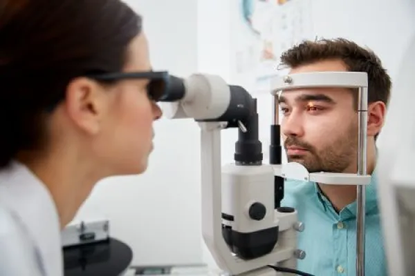
For corneal FBRs, CPT® instructs coders to use 65220 (… corneal, without slit lamp) or 65222 (… corneal, with slit lamp) depending on slit lamp use. “The slit lamp is a microscope that emits a focused narrow, high-intensity beam of light. It may be used when examining the eye to find an FB or to identify abrasions or other injuries to the eye caused by an FB,” notes Linda Martien, COC, CPC, CPMA, CRC, of Medical Revenue Cycle Management Consulting.
Lempert reminds coders that the physician might also use a slit lamp for conjunctival FBs, but there is no separate CPT® code for the use of a slit lamp on these FBRs. “A slit lamp is used to get a better view of the eye so that a precise method can be used to remove an FB. It allows the clinician to see the eye and FB much better,” explains Lempert.
The physician can decide to use — or not use — a slit lamp for a variety of reasons. “Sometimes it is a personal choice whether to use a slit lamp or not. It depends on the patient’s cooperativeness, the clinician’s confidence, the FB in question, the exact location of the FB, the availability of a slit lamp … and other factors,” Lempert says.
Examples: Lempert offers two scenarios; one illustrates when a slit lamp is used and one when it’s not used. “An example of 65220 is an FB on the cornea that can be removed with a Q-tip. An example of 65222 is an FB that will not come out with a Q-tip because it is stuck and needs to be removed by a scoop and picked at for a while before it will move and can be removed.”
Consider This Clinical Example
For a more detailed FBR encounter, consider this example from Martien:
A patient presents with a complaint of pain in the right eye, with tearing and burning. The patient was working in the yard pruning and trimming bushes earlier today. They were not wearing protective eye covering and think a piece of grass or other debris flew into their eye. The patient does not use any eye medications.

Examination shows the patient’s visual acuity is 20/40 in the left eye and 20/20 in the right eye. Intraocular pressures are within normal limits. There is no nystagmus. The right eye is dilated, and the slit lamp is brought into position. There is a 2 mm x 2 mm corneal epithelial defect (+fluorescein staining) and a very small object, apparently, a grass seed, is seen. There is no corneal stromal infiltrate or thinning. The seed has most likely scraped the cornea, causing a minor abrasion.
A moistened cotton swab is used to successfully remove what is, as suspected, a small grass or plant seed. Antibiotic eye drops are prescribed for the patient to use at home over the next seven days.
For this claim, you’ll report:
Warning: Martien says you should not provide a diagnosis code for corneal abrasion. According to the American College of Ophthalmology, it may cause confusion and result in a claim denial. (www.aao.org/practice-management/news-detail/foreign-body-with-abrasion-noted).
