Medical Coding’s Best Online Code Search & Lookup Resource
What's New?
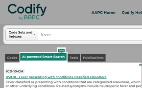
AI-powered Smart Search bar visibility continuous at the top of the page.
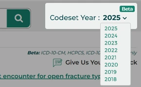
The Historical Code Set feature reverts Codify to previous years, displaying only the code set data relevant to the chosen date or quarter.

Custom-arrange the tools and boxes on your main tool page and code detail pages

Easier, top-menu access to publications included in your accounts


RHIT, CRC – Auditor

Codify Your Team!
Reduce denials and maximize revenue with our most powerful encoder.
Request A QuoteStart Your Free Trial Of Codify by AAPC Today!
Coder Search
$10/mo
(Non-Members $15.00)
Basic Coder
$36/mo
(Non-Members $48.00)
- Includes Coder Search
- NCCI Edits
- NCD & LCD Lookups
- Crosswalks
Pro Fee Coder
$44/mo
(Non-Members $57.00 )
- Includes Basic Coder
- Historical Code Sets
- CMS 1500 Real-Time Scrubber
- Revenue Cycle Insider Access
- Specialty Survival Guide
- 10 Optional Add-ons
- 20 CEUs/Year
Complete Coder
$54/mo
(Non-Members $66.00)
- Includes Pro Fee Coder
- OPPS CCI Edits Checker
- DRG Lookup
- ASC Lookup
- ICD-10-PCS Code Lookup
- 12 Optional Add-ons
- 20 CEUs/Year
Codify by AAPC Product Comparison
Coder Search
Basic Coder
Pro Fee Coder
Complete Coder
Essentials
Codify to eBook Integration feature enables subscribers to effortlessly access and navigate their eBook content by clicking the eBook icon, while also allowing them to conveniently access specific code snippets in Codify by selecting them in the eBook.
Clean intuitive page design with drag/drop widgets for dashboard and code details customization
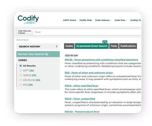
Universal Search for code sets, indexes, tools, publications
AI-powered Codify Smart Search
Official descriptors with guidelines, lay terms, illustrations, BETOS, and date-related information
Every coder has the essential tools needed to perform their coding as efficiently as possible. Codify provides those tools to you — including ICD-10-CM cross references, E/M calculator, AMA CPT® Section Guidelines, global surgery days calculator, and personal notes option so you keep track of knowledge learned from payers or other sources.
Store commonly used and favorite codes
Current and upcoming code changes
Common crosswalks — ICD-9 to ICD-10 and CPT®/ HCPCS to BETOS
Superbill Converter
Codify’s Publication Library contains multiple publications from CMS, Federal Register, Medicaid, OIG HSS, private payers, and state fraud control websites.
Annual/historic specialty CPT®/HCPCS/ICD-10 code changes
In App just-in-time tutorials and FAQs
Newsletter Library (20 CEUs)
Tools & Fee Schedules
The E/M Calculator guides you to the right code based on your date of service. You’ll find 1995 and 1997 Documentation Guidelines, as well as 2021, 2023, and the latest updates in one tool.
MPFS for facility and nonfacility
Modifier Guidelines ASC and P (Ambulatory Surgery Center and Physician)
NCCI Edits Checker with Change Version and Medicaid NCCI edits Checker
The Part B Physician Fees & More widget allows you to look up the fee schedule for a code based on the year and quarter. The results — from one simple search — provide you not only with the fee schedule but also with the RVU data, global surgery days, modifier guidelines, and MUEs.
Codify contains 18 different crosswalks to assist you in mapping one code set to another or from code to modifiers, revenue codes, LCD/NCD’s and more.
Revenue code lookup and PQRS (MIPS)
Advanced Tools & Options
CMS 1500 Real-Time Scrubber
The Historical Codeset feature allows you to access historical medical coding data for a specific period by reverting Codify to previous quarters, displaying only the code set data relevant to the chosen date or quarter.
Specialty Survival Guide
Access to the Coder Chat Forum* (Membership required)
Unlimited claim entry into Claim Scrubber
OPPS NCCI edits Checker
DRG to IPPS/LTCH Crosswalk
OPPS & ACS Fee Schedules

Frequently Asked Questions
Have a different question? Submit inquiries through our contact us page.
Codify is our newest revenue cycle platform to allow subscribers the fastest tool for code look-up among multiple additional tools and services.
Codify’s data is updated regularly to ensure the information provided to you is as accurate as possible. CPT® and ICD-10 codes are updated annually, and HCPCS Level II codes and NCCI edits are updated quarterly.
Google Chrome and Mozilla Firefox works best. Internet Explorer and Edge can cause some unexpected results.
The number of administrators for Codify is unlimited, just like it was with AAPC Coder.
To log in to Codify, go to AAPC.com and log in to your account, click on My AAPC or Resources from the top banner. Select Codify from the options available.
Codify licenses are assigned individually, which means only one individual can access Codify with login credentials. If someone tries to access Codify with your account information, you will forfeit access to the program will be kicked out of the program.
Codify is a web-based program, so you can use it on any device with internet access.
Codify offers the ability to earn up to 20 CEUs annually by logging in and reading through the Survival Guides and completing Quizzes under the CEU tab.
Codify offers multiple add-on features based on the product you purchased. Please see the Add-on page for more details.
Yes, you can look back at past fee schedules, code changes, and any historical information that has changed per code.
Codify has past fee schedules, code changes (including New, Revised, and Deleted code lists), LCD/NCD data, articles, coding clinics, regulatory publications, E/M guidelines, and NCCI or NCCI edits. The Symbols data is not included in this information.
AAPC provides training and assistance for businesses to ensure all users can use the platform to its fullest capacity. Our training also walks administrators through the admin functions of Codify.
Once your account is activated you will receive a welcome email with your login credentials and instructions on how to access Codify.
AAPC Coder and SuperCoder were updated to provide the best platform available to our subscribers. This update provides more functionality for medical billing and coding staff.
Yes, once you log into Codify your favorites and notes will automatically merge into Codify. If you choose to toggle between the two platforms prior to closure of the original platform, your notes and favorites will auto-update again to ensure all your data is current and up to date.
Codify is not refundable, you may terminate your subscription or free trial by contacting AAPC via phone, live chat, or email. To ensure that your credit card does not get charged, please make your cancelation request is at least two business days prior to the end date of your subscription or free trial term.
CPT® is a registered trademark of the American Medical Association